Infrared
Photography and Double Helix Water
We not claim health
cures, however our research and testimonies point to the idea
that drinking
even a small amount of stable water cluster may positively effect health
and healing.
Experiments
Using InfraRed Photography
(Please visit these
pages for sample images.)
Infrared Photos,
before and after photos, after 15 minutes of drinking SWC.
Arthritis
Autism
Spectrum Disorders
Fibermyalgia
Hand
Migraines
Male Page
Women’s
Page
Thyroid
What is Infra Red Photography?
Medical DITI is
a noninvasive diagnostic technique that allows the examiner to visualize
and quantify changes in skin surface temperature. An infrared scanning
device is used to convert infrared radiation emitted from the skin
surface into electrical impulses that are visualized in color on a
monitor. This visual image graphically maps the body temperature and
is referred to as a thermogram. The spectrum of colors indicate an
increase or decrease in the amount of infrared radiation being emitted
from the body surface. Since there is a high degree of thermal symmetry
in the normal body, subtle abnormal temperature asymmetry’s can be
easily identified.
Skin blood flow
is under the control of the sympathetic nervous system. In normal people
there is a symmetrical dermal pattern which is consistent and reproducible
for any individual. This is recorded in precise detail with a temperature
sensitivity of 0.1°C by DITI. The neurochemistry application of
DITI measures the somatic component of the sympathetic nervous system
by assessing dermal blood flow. The sympathetic nervous system is stimulated
at the same anatomical location as its sensory counterpart and produces
a ‘somato sympathetic response’. The somato sympathetic response appears
on DITI as a localized area of altered temperature with specific features
for each anatomical lesion. The mean temperature differential in peripheral
nerve injury is 1.5°C. In sympathetic dysfunction’s (RSD / SMP / CRPS)
temperature differentials ranging from 1° C to 10° C depending on severity
are not uncommon. Hematological processes generally appear as ‘hot’
areas with increased temperature patterns. The pathology is generally
an inflammatory process, i.e. synovitis of joints and tendon sheaths,
epicondylitis, capsular and muscle injuries, etc. Both hot and cold
responses may coexist if the pain associated with an inflammatory focus
excites an increase in sympathetic activity. Also, vascular conditions
are readily demonstrated by DITI including Raynauds disease, Vasculitis,
Limb Ischemia, DVT, etc. Medical DITI is filling the gap in clinical
diagnosis … X ray, C.T. Ultrasound and M.R.I. etc., are tests of
anatomy. E.M.G. is a test of motor physiology. DITI is unique in its
capability to show physiological change and metabolic processes. It
has also proven to be a very useful complementary procedure to other
diagnostic modalities. Unlike most diagnostic modalities DITI is non
invasive. It is a very sensitive and reliable means of graphically
mapping and displaying skin surface temperature. With DITI you can
diagnosis, evaluate, monitor and document a large number of injuries
and conditions, including soft tissue injuries and sensory/autonomic
nerve fibre dysfunction.
There is growing
use of thermography for early breast cancer detection. American
College of Clinical Thermology
Thermography as a diagnostic tool is not
universally accepted as a diagnostic tool, however we are using
it as an indicator
that there has been change. We are not using infrared photography to
identify illnesses.
Following images is from a subject with
severe computer hand injury.
Computer Hand
Injury
Pre and Post infrared images of a subject
with long term computer hand injury. Her first image
did not show fingers, it is speculated that her circulation in her hand
was poor. Subject drank
2 8 oz glasses of Stable Water Cluster for two weeks. Her
post images
showed all her fingers and a more even and healthier distribution of
color.
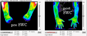
Back of hand view pre SWC and Post SWC
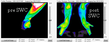
Side of hand view pre SWC and Post SWC
Dr. Lo is guessing that there is a movement
towards health
when the images move from not showing fingers, to showing
all fingers and moving towards a more
evenness in temperature.
Go to top to see infrared experiments
stablewatercluster.net
all rights reserved ©2010
|